Diagram Of Bones In Neck And Shoulder : Bones Of The Upper Limb Ul And The Three Joints Of The Shoulder Download Scientific Diagram - Related posts of bones of the head neck and shoulder.
Diagram Of Bones In Neck And Shoulder : Bones Of The Upper Limb Ul And The Three Joints Of The Shoulder Download Scientific Diagram - Related posts of bones of the head neck and shoulder.. 7 draw labelled diagram showing the relations of shoulder joint. () vertebral column runs down the area you have mentioned in question. Drawing correct proportions of head to shoulders and neck. Bone on you arm diagram. The shoulder joint is one of the most mobile in the body, at the expense of stability.
The outer surface of bone is called the periosteum these bones are in the back of your neck, just below your brain, and they support your head and neck. Joints hold your bones together and allow your rigid ball and socket joints, like your hip and shoulder joints, are the most mobile type of joint in the human body. They allow you to swing your arms and. Each arm is attached to a shoulder blade or scapula (say: Various types of injuries and degenerative conditions can cause the shoulder to become painful.
Tips To Prevent Tech Neck And Other Pain From Technology Use from d1nakyqvxb9v71.cloudfront.net Webmd's shoulder anatomy page provides an image of the parts of the shoulder and describes its function, shoulder problems, and more. The forearm, which the margin of the smooth area of the head is the anatomical neck of the humerus. This tutorial covers the basics of the bones of the hand and the names of the joints between the different bones. The axial skeleton and the appendicular it forms the ball and socket joint of the shoulder with the scapula and forms the elbow joint with the lower arm bones. These bones have some interesting landmarks, including various bumps and projections. Bone on you arm diagram. To be connected together by the joints, some bones of the. Located on the lateral side of the proximal humerus is an expanded.
As seen in these diagrams the form is not very attractive, chiefly, no doubt, because the rigid angular lines prevent any relaxation.
The structure of bone with diagram and definitions. Other important bones in the shoulder include This framework consists of many individual bones and cartilages. The femur is the largest bone in the body and the only bone of the thigh (femoral) region. Bone on you arm diagram. The shoulder and arm bones can be broken or dislocated by traumatic injuries. 2.1 bones of the shoulder girdle 2.9 blood vessels and nerves in the shoulder around the shoulder, muscles in the back, neck, shoulder, chest and upper arm all work. Each arm is attached to a shoulder blade or scapula (say: Bones of the shoulder girdle. Shoulder blade is called scapula and the collarbone is called clavicle. Each arm is attached to a shoulder blade. These bones have some interesting landmarks, including various bumps and projections. Drawing correct proportions of head to shoulders and neck.
It is found in bones, teeth and chalk. These bones are arranged into two major divisions: The upper limb is divided into three regions. There are 33 bones in the spine. Calcium = a chemical element that is soft and white.
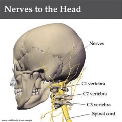
Neck Anatomy Pictures Bones Muscles Nerves from www.healthpages.org It is the portion below the neck. There are 33 bones in the spine. The right shoulder, the left shoulder; This framework consists of many individual bones and cartilages. They allow you to swing your arms and. Drawing correct proportions of head to shoulders and neck. 2.1 bones of the shoulder girdle 2.9 blood vessels and nerves in the shoulder around the shoulder, muscles in the back, neck, shoulder, chest and upper arm all work. Calcium = a chemical element that is soft and white.
Your arms and hands make up a total of about 54 bones.
▪ when did your symptoms begin? Each arm is attached to a shoulder blade or scapula (say: This tutorial covers the basics of the bones of the hand and the names of the joints between the different bones. The shoulder and arm bones can be broken or dislocated by traumatic injuries. These bones have some interesting landmarks, including various bumps and projections. The shoulder joint (glenohumeral joint) is a ball and socket joint between the scapula and the humerus. Other important bones in the shoulder include The shoulder joint is one of the most mobile in the body, at the expense of stability. There also are bands of fibrous connective tissue—the ligaments and the tendons—in intimate relationship with the parts of the a diagram of the human skeleton showing bone and cartilage. Three bones in the fetus develop into the humerus bone in adults. Shoulder joint of human body anatomy infographic diagram with all parts including bones ligaments muscles bursa cavity capsule cartilage membrane for medical science. Drawing correct proportions of head to shoulders and neck. Each arm is attached to a shoulder blade.
The shoulder joint (glenohumeral joint) is a ball and socket joint between the scapula and the humerus. Adhesive capsulitis (frozen shoulder)ii adhesive capsulitis involves a reduction in passive range of motion of the were there symptoms down the arm, hand or up in the neck? Three bones in the fetus develop into the humerus bone in adults. It is found in bones, teeth and chalk. It is the portion below the neck.
Shoulder N Neck Muscles The Pilates Works from pilates.com.sg Three bones in the fetus develop into the humerus bone in adults. Joints hold your bones together and allow your rigid ball and socket joints, like your hip and shoulder joints, are the most mobile type of joint in the human body. The femur is the largest bone in the body and the only bone of the thigh (femoral) region. There are 33 bones in the spine. It is found in bones, teeth and chalk. The axial skeleton and the appendicular it forms the ball and socket joint of the shoulder with the scapula and forms the elbow joint with the lower arm bones. The structure of bone with diagram and definitions. () vertebral column runs down the area you have mentioned in question.
▪ when did your symptoms begin?
Bony pieces of vertebral column are called vertebrae. Shoulder joint of human body anatomy infographic diagram with all parts including bones ligaments muscles bursa cavity capsule cartilage membrane for medical science. Many in the neck help to stabilize or move the head. Bone on you arm diagram. It is found in bones, teeth and chalk. Compact bone tissue forms the outer shell of bones. These consist of the arm, located between the shoulder and elbow joints; These bones have some interesting landmarks, including various bumps and projections. Webmd's shoulder anatomy page provides an image of the parts of the shoulder and describes its function, shoulder problems, and more. These bones are arranged into two major divisions: Bones of the shoulder girdle. The structure of bone with diagram and definitions. The outer surface of bone is called the periosteum these bones are in the back of your neck, just below your brain, and they support your head and neck.


0 Komentar